Dr. Sinha[1] is the anti-Benjamin of our times. He asks his patients to believe in the aura of the machine. When I meet him in the treatment room at the corner of the Radiation Oncology wing on a Thursday afternoon—its ceiling painted blue with drifting, improbable clouds—he speaks in paragraphs, waxing lyrical about the CyberKnife’s precision. The CyberKnife’s industrial robotic arm, he tells me, is a direct import from the automobile assembly line. At his cue, the technologists in the control room set it in motion. The arm whirrs as it moves along three linear and rotational axes; it is designed to track the movements of the patient’s body in real time. Dr. Sinha walks me through each piece of the ensemble: the ceiling-mounted x-ray machines that track the tumour’s position, the large linear accelerator that charges the radiation beam, a phantom skull laid on the table for setting delivery coordinates. “High dose and minimal margins,” he insists, “is the future of radiation therapy.”
One cannot help but be convinced that this is what medical salvation looks like. In some respects, I am already a believer. In May 2025, a month after receiving a stage IV lung cancer diagnosis, I underwent image-guided radiation therapy under Dr. Sinha’s supervision, becoming one of the very few patients approved for this advanced form of stereotactic radiosurgery. After months of misdiagnosis from pulmonologists working through experience and statistical generalisation, it was computer-aided imaging that revealed the adenocarcinoma that was rapidly spreading through my body. By looking at the MRI image, any oncologist could tell that my cancer had metastasized into the brain, creating eight lesions that could not be targeted through traditional chemotherapy. At this stage, even radiation therapy was, to borrow Julie Livingston’s words, “a necessary exercise in hope” (2012: 161).
As I underwent radiation therapy under the CyberKnife, I began compiling field-notes on my encounters with the radiosurgery system. They were fragmented autoethnographic vignettes that located the CyberKnife and its tacit promise of immortality within the immediate rupture left open by the cancer diagnosis. In June, I wrote a short article in Somatosphere that weaved together these reflections. My analytic aperture was the discursive regime surrounding the machine: how precision was staged rhetorically against uncertainty, how the aura of high technology transformed the patient’s relation to their own finitude, and how the claims of biomedicine fit within a broader cultural milieu of technological solutionism.
In the following months, however, I saw the promise of the CyberKnife from other perspectives. As my cancer began responding to therapy, I found myself in an introductory workshop to the CyberKnife, being walked through the system’s workflow alongside a group of medical students. A few months later, I worked with a media organisation to document the system for public awareness, interviewing the radiation oncology team and filming the machine in action. Across these encounters—as patient, social scientist, and journalist—stereotactic radiosurgery revealed itself as something more than a reified fantasy of life. It opened onto an assemblage of practices that rendered the body into data, a site where the climactic expression of precision medicine was staged alongside a carefully orchestrated spectacle of medical futurity.
In this piece, I build on my earlier exploration of the CyberKnife to interrogate the image of pathology that anchors this formation. In the CyberKnife, the image ceases to be an artefact that represents certain facts about the diseased body. As it is folded into the infrastructural substrate of computational planning, the image transforms from an inert object that offers a perspectival view of pathology into a malleable data cluster that redefines the nature of radiosurgical action. This collapse between image and event, I hope to show in this article, reconfigures the practice of modern radiosurgery. What emerges from this collapse is a new kind of medical machine, where the image moves from the surface of the screen to the innards of algorithms.
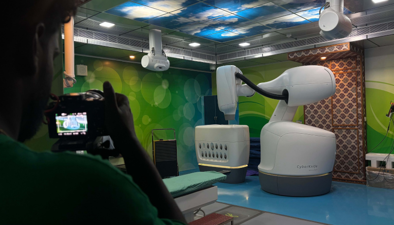
A cameraperson from the film crew records the CyberKnife in action. Photo by the author.
Image as a Representative Object
In biomedicine, image has long served as a “substitute for bodies” (Saunders 2008: 14), a representational device that enabled the clinician to see the diseased body with greater fidelity. From X-rays to computed tomography (CT) images, successive advances in imaging have expanded both the morphological resolution and the perspectival diversity with which the body can be apprehended by the clinician. Today, the ocular capacity of a hospital is distributed across a meshwork of machines that continuously generate and circulate images through the hospital’s PACS—the picture archiving and communication system. From the moment a patient is admitted, devices such as echocardiograms, video laryngoscopes, ultrasound scanners, CT machines, and positron emission tomography (PET) scanners produce a steady stream of images: an economy of visual fragments that are woven together to inform clinical decision-making.
Yet, the reproduction of the body as image is anything but straightforward in modern biomedicine. As studies of medical imaging and digital diagnostics have shown (Beaulieu 2002; Saunders 2008), the body is increasingly encountered in clinical settings today through layers of computational abstraction, where visibility is achieved through calculative operations rather than direct observation. For instance, in his pioneering work on magnetic resonance imaging (MRI) machines, Amit Prasad calls attention to how mathematical and geometrical techniques become central to measuring pathology, where probabilistic body atlases form the foundation of establishing normality and abnormality. As Prasad notes, MRI machine makes the body notational, allowing the radiologist to look at image fragments and “visually extract” pathology from a body part (Prasad 2005: 293).
Inserted into a vast infrastructure of medical gaze, images also move rapidly to form a “cross-referential” network of diagnostic and clinical tools (Prasad 2005: 300) that is accessible by any doctor in the hospital. Their points of origin cease to matter. As they coalesce into a diagnostic aggregate, a clinician can extract any detail (three sizeable lesions, a flask-shaped accumulation of pericardial fluid, cloudy shadows in the lung) to make specific conclusions that align with their expertise. A single image, such as a chest x-ray, is only one segment in a sequence of images that maps the progression of a disease. These images remain fundamentally representative: instruments for longitudinal comparison, used to stabilise and contrast the state of the body as biomedicine acts upon it over time.
Ways of Acting Algorithmically
My encounters with the CyberKnife suggest that computer-aided radiosurgery cannot be understood as a lineal descendant of the cyborg visuality theorised by Amit Prasad over two decades ago. It represents a radical shift in the essentials of radiosurgical practice: the ways in which the CyberKnife intervenes on pathology—its ways of visualising the tumour as actionable coordinates, its capacity to continuously reorient itself to the liveliness of the human body, and most critically, its ability to capture the imagination of those who interface with it—transforms image from a modality of representation to a modality of intervention.
CyberKnife treatment—like other form of three-dimensional (3D) radiotherapy—begins with CT simulation, a process where a CT scanner is used to generate a 3D image of the tumour. This spatialises the tumour into something the machine can recognise as a target. This is followed by contouring, a process in which oncologists work with radiologists, dosimetrists, and medical physicists to delineate target volumes on the image of the tumour. At this stage, the team defines the margins around the tumour and accounts for motion and setup errors. The team may also employ supplementary, software-based techniques such as image fusion to combine different imaging modalities for better tumour definition. The conventions of oncological labour here are designed not to discover certain facts about the tumour, but to define the tumour graphically as a stable, operable object.
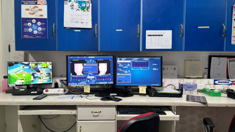
Control room for the CyberKnife radiation therapy system. Photo by the author.
During treatment delivery, however, the CyberKnife appropriates these representative conventions, if only to subvert them. CyberKnife’s proprietary algorithms use images—data from digitally reconstructed radiographs, ceiling mounted X-ray imagers, and optical cameras in the treatment room—to translate the body into live informational coordinates. The image of pathology carefully created by the tumour board is also absorbed into this formation, becoming something more than a planning aid for the human eye. For treatment delivery, the CyberKnife relies on inverse planning algorithms, which work backwards from desired clinical outcomes to compute thousands of possible non-isocentric beam trajectories[2] . They allow the machine to algorithmically model treatment—deciding the angles from which radiation beams must be aimed at the tumour, the dose with which a structure must be bombarded, and how the robotic arm must compensate for the movement of the patient’s body.
This makes the CyberKnife decidedly different from older imaging modalities such as MRI. As Prasad observes, diagnostic certainty in medical imaging has historically been attempted by combining a differential analysis of images along with other diagnostic inputs. For instance, a radiologist who is performing a CT angiogram might be “looking” for a specific anomaly depending on the notes provided by the medical oncologist. In the CyberKnife, however, the image is more than a representation to be acted upon by human judgement. The CyberKnife’s promise of precision—enabled by its capacity to “see” the tumour algorithmically—is not burdened by an attempt to arrive at a diagnostic truth. Rather, representation of pathology is merely a layer within a continuous feedback loop through which human expertise, hardware, and computation converge in radiosurgical action.
A Monument of Medical Salvation
The CyberKnife inaugurates a visual regime where the re-rendering of the body as information not only informs decisions made by a physician, but also affords significant degrees of automation in the process of radiosurgery. As the machine undoes the distinction between observation and action, human expertise recedes behind its charisma, demanding that the spectacle of medical salvation be staged in other ways.
This becomes apparent when I meet Dr. Sinha for the shoot. What intrigues me during our conversation is his investment in the atmospherics of the machine. To the camera crew, he insists on a slow push-in, the CyberKnife easing into the shot. He wants its feel captured: the control room with rows of monitors and revolving chairs, the collimator changer with twelve cones arranged symmetrically, the imposing robotic arm of the CyberKnife gliding against the crescent moon in the ceiling sky. He wants the viewer to sense what precision medicine looks like. He wants a work of art in the age of mechanical reproduction.
Dr. Sinha’s commitment to this spectacle of medical modernity reminds me, yet again, of what Ernest Becker (1973) suggested in his meditation on mortality. If we are, as Becker suggests, always suspended between our awareness of death and our attempts to deny it, technologies like the CyberKnife offer a particularly compelling language for that denial. And perhaps that is where their power lies—not in overcoming death, but in making it possible to imagine, however briefly, that it can be managed through the charisma of technology.
Notes
[1] Name changed
[2] In the CyberKnife system, the radioactive beam can be directed from any angle.
This post was reviewed by Contributing Editor Ritu Ghosh.
References
Beaulieu, Anne. 2002. “Images Are Not the (Only) Truth: Brain Mapping, Visual Knowledge, and Iconoclasm.” Science, Technology, & Human Values 27(1): 53–86. https://doi.org/10.1177/016224390202700103
Becker, Ernest. 1973. The Denial of Death. New York, NY: Free Press.
Livingston, Julie. 2012. Improvising Medicine: An African Oncology Ward in an Emerging Cancer Epidemic. Durham, NC: Duke University Press.
Prasad, Amit. 2005. “Making Images/Making Bodies: Visibilizing and Disciplining through Magnetic Resonance Imaging (MRI).” Science, Technology, & Human Values 30(2): 291–316. https://doi.org/10.1177/0162243904271758
Saunders, Barry. 2008. CT Suite: The Work of Diagnosis in the Age of Noninvasive Cutting. Durham, NC: Duke University Press.
Chakramakkil, Thomson. 2025. “Immortality in the Machine: Autoethnographic Notes from Radiation Therapy.” Somatosphere. https://somatosphere.net/immortality-in-the-machine-autoethnographic-notes-from-radiation-therapy/

1 Comment
I wish you a good day, and less pain.